

Johns Hopkins University Campus in Baltimore, Maryland. It was founded on January 22, 1876, and named for its benefactor, the philanthropist Johns Hopkins. The institution pioneered the concept of the modern research university in the United States and has ranked among the world's top such universities throughout its history.

The National Science Foundation (NSF) has ranked Johns Hopkins #1 among U.S. academic institutions in total science, medical and engineering research and development spending for 31 consecutive years. As of 2011, thirty-seven Nobel Prize winners have been affiliated with Johns Hopkins in a span of just 135 years, and the university's research is among the most cited of any institution globally, making it one of the most prestigious universities in the world.

The campus is constructed in the style of Federal Architecture. Federal-style architecture is the name for the classicizing architecture built in North America between c. 1780 and 1830, and particularly from 1785 to 1815. The style broadly corresponds to the middle-class classicism of Biedermeier style in the German-speaking lands, Regency style in Britain and to the French Empire style.

The University hosted Johns Hopkins CTY program seminar on Neurology. Concepts covered by the seminar included Neuroanatomy, Exploring how the Brain works, Signals into and Out of the Brain: Turning Thoughts into Action, Attention & Awareness: Focusing on What Matters, Sensory Illusions: Why you can't always trust your senses. The seminar included lectures, laboratory exercises and work-shops.

We were assigned into the Temporal Lobe Team, and were covering Attention and Awareness here in the Undergraduate Teaching Lab.

Neural mechanism of attention and awareness. There are two sources in the brain from which an attentional signal originate: 1. The prefrontal lobe provides "top-down" signal to help us to direct our attention according the goals that we set for ourselves. 2. The sensory cortices provides relay newly incoming sensory information to and direct our attention to the significant and most salient stimuli in the world. However, sometimes these two signals may conflict with each other, causing inattentional and change blindness.

Neuroimaging - exploring human brain. One of the most interesting ways that we can study neuroscience is by looking at the brain. However, it's very difficult to study the human brain originally. Several groundbreaking techniques that neuroscientists have used are PET (Positron Emission Tomography) as well as fMRI (functional Magnetic Resonance Imaging) and DTI (Diffusion Tensor Imaging).

Neuroanatomy - looking at the sections and functions of brain.

Exploring anatomical behavior in the brain. Daniel is looking at dendrite in a neuron of a mouse. When a signal travels from one neuron to another neuron, the electrical charge is converted into a chemical messenger system between the sending neuron's axon and the receiving neuron's dendrite. These dendrites have small spikes all over them, called dendritic spines. Dendritic spines are little compartments that boost the incoming signal independently and then send their boosted signal to the receiving neuron's soma. These spines are important, because they allow for more computational power per input. That means they are especially useful in learning and memory formation.

When we examine what part of the brain is important for memory, we're looking here at behavioral data. Graduate students explain here how mice store memory in their cortex part of the brain through a maze experiment.

Testing body based illusions. Here the experiment confuses the testers brains by providing discordant visual, tactile and proprioceptive stimuli. When the brain gets information that does not perfectly match up, it tries to construct a coherent story. This tricks the body into adopting something that may not be real.

Tactile illusions work-shop. The somatosensory system is involved in our perception and sensation of touch. The skin contains at least three major types of receptors: Merkel Receptor - light touch, Pacinian Corpuscles - Deep touch and pressure and Ruffini Corpuscles - stretching and temperature. Jasmin and Daniel are about to experiment with different ways the brain alters perception.

The Aristotle Illusion: While blindfolded, the testers are asked to hold out their hands while having their index and middle finger crossed. An object (here a pen) will touch the junction of the crossed fingers. Many will feel two objects touching them. This is an example of object disjunction.

Lunch break time at the Levering Hall!


Enjoying the campus on lunch break. Red Maple in her full glory here!!

Jasmin and Daniel agreeing to stand still for a picture. :)

My camera lens seems to be completely infatuated with the colors of the season. :)

Entering into the brand new Undergraduate Teaching Laboratories as the very first visitors.

Project Lab G72 and one of the territories of the Postdoctoral Fellow Dr. Sam Nummela. Yes, he has finnish ancestors. :)

This laboratory explored how neurons encode and transmit sensory signals from receptors to the brain and motor commands from the brain to muscles. This knowledge is applied to understand the principles behind neural prosthetics.

Dr. Sam Nummela demonstrating how the nervous system of the America cockroach can be manipulated via stimuli from human brain.

Daniel is a tester here. He has electrodes attached to his scalp which are also attached to a C-ISO amplifier. A freshly dissected cockroach leg is also attached into the amplifier. The signal which Daniel sends to his arms when commanding them to move, is also sent to the cockroach's leg, making it move too. View a video of this inspiring exercise below!
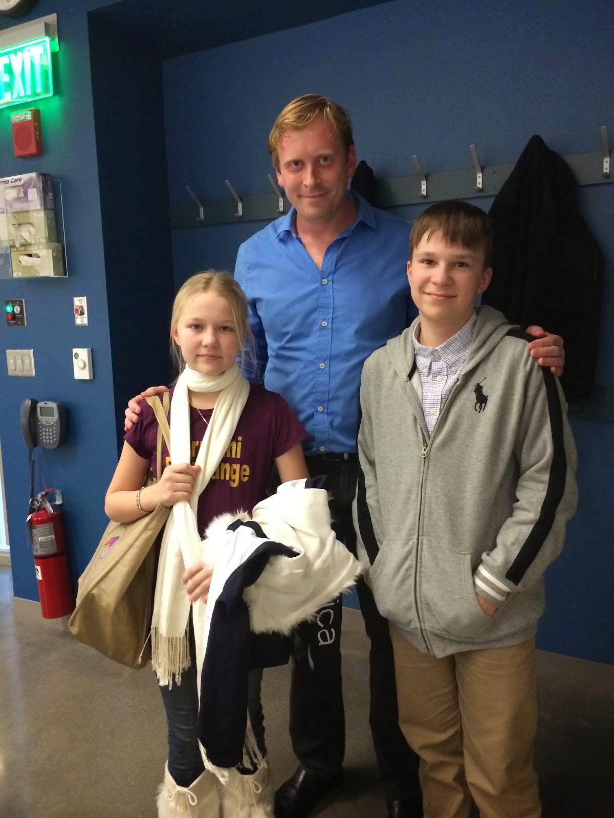
Jasmin and Daniel enjoying a visit with Dr. Sam Nummela.

Gorgeous views of the campus area from the floor-to-ceiling windows of the Lab.

Neurology graduate students of the Johns Hopkins University share some of their knowledge with the young "apprentices".

Time for dissections! This is a brain of a sheep.

Jasmin is being assisted by a Neurology student from the Johns Hopkins to find the different sections of the brain.

Toivo and Daniel are doing the same with their assistant.

The Medulla and Cerebellum have been cut out.

A close encounter with real human brain. Daniel was the lucky chosen student who got to handle this prestigious property of the John's Hopkins Neuroscience lab.

The fabulous Dr. Gorman explaining more details about the specimen. It belonged to a 67-year old (healthy) male from Michigan.

Feeling grateful and staying in the premises after an inspiring day... :)

Evening setting in at the Johns Hopkins University campus.
No comments:
Post a Comment